Spine Conditions & Procedures
Lancaster NeuroScience & Spine Associates

Providing comprehensive treatment for a variety of neck and spine-related conditions
About our Diagnosis and Procedures
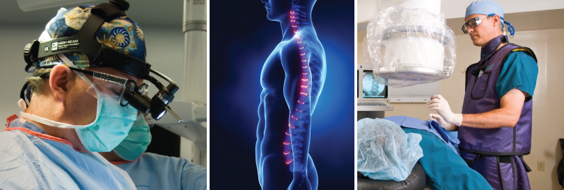
Our team of Neurosurgeons and Physiatrists treat a variety of neck and spine-related conditions including those listed below. Our evaluation process lends treatment options that include Physical Therapy, non-invasive procedures and surgery, all of which we provide compassionate care from diagnosis to recovery.
Commonly treated Spinal Conditions (Neurosurgery)
- Herniated Disc
Herniated Disc
The disc is a soft, spongy-like material that both cushions and supports the bones of the spinal column, called vertebrae. At times, the disc can herniate or protrude out of its normal lining. When this happens, the disc can pinch upon a nerve or upon the spinal cord itself, and cause a variety of symptoms.
Procedures used to treat Herniated Disc
- Laminectomies & Spinal Fusions
- Spinal Stenosis
Spinal Stenosis
The term spinal stenosis refers to a narrowing of the spinal canal, the area where the spinal cord and nerve roots run. When severe, spinal stenosis can cause significant pain in the legs and difficulty walking over prolonged periods. Many treatment options currently exist, and may be discussed with your surgeon.
Procedures used to treat Spinal Stenosis
- Laminectomies & Spinal Fusions
- Vertebral Compression Fracture
Vertebral Compression Fracture
A vertebral compression fracture (VCF) is a break in the vertebrae that causes it to collapse. This causes the spinal column above it to bend forward. Most VCF’s occur in people with underlying osteoporosis, although these fractures can also occur without a specific cause. They may cause significant back pain that is unresponsive to rest or pain medications. They can lead to serious health problems, including eating and sleeping disorders, lung conditions, and difficulty walking or performing normal activities.
Procedures used to treat Vertebral Compression Fracture
- Kyphoplasty
- Spinal Instability
Spinal Instability
Spinal instability refers to the inability of the spinal column to support itself. There may be excessive motion, which contributes to a pinching of a nerve or narrowing of the area where the nerve runs. In general, the causes of this are quite varied.
Procedures used to treat Spinal Instability
- Laminectomies and Spinal Fusions
- Spinal Cord Tumors
Spinal Cord Tumors
Thankfully, spinal cord tumors are generally rare. There can be both malignant and benign spinal cord tumors. Neurosurgeons specialize in the diagnosis and treatment of tumors in the brain and spinal cord. The symptoms of a spinal cord tumor generally depend on its size, rate of growth, and location.
Procedures used to treat Spinal Cord Tumors
- Laminectomies & Spinal Fusions
- Spinal Injuries
Spinal Injuries
Injury to the spinal cord or to the nerves can be quite devastating. Neurosurgeons specialize in the care of this type of injury. There are, unfortunately, few treatments to help the nerves or spinal cord regenerate. However, much research is being done in this area.
Procedures used to treat Spinal Injuries
- Laminectomies and Spinal Fusions
Routinely Performed Spinal Procedures (Neurosurgery)
- Laminectomies & Spinal Fusions
Laminectomies & Spinal Fusions
Spinal operations are performed to decompress neural structures to relieve pain and provide the opportunity for neurological recovery. At times, fusions are performed to stabilize spinal segments and improve neurologic function. These procedures can be peformed microscopically, endoscopically, or through standard, open approaches.
Conditions routinely treated with Laminectomies & Spinal Fusions
- Herniated Disc
- Spinal Stenosis
- Spinal Instability
- Spinal Cord Tumors
- Spinal Injuries
- Kyphoplasty
Kyphoplasty
Surgeons perform a minimally invasive procedure, called kyphoplasty, to treat painful compression fractures of the vertebrae of the spine. It is performed by inserting an inflatable balloon through a very small incision. A small tube is inserted down into the bone to create a path into the fracture. The balloon is inserted and inflated, creating a space that can be filled with bone cement. Kyphoplasty has a very high success rate for pain relief, fracture reduction and stabilization.
Conditions routinely treated with Kyphoplasty
- Vertebral Compression Fracture
Commonly treated Spinal Conditions (Physiatry)
- Sciatica
Sciatica
The sciatic nerve supplies sensation to the posterior thigh and buttock, knee flexors, and foot muscles. When this nerve is compressed, inflamed, or irritated anywhere along its length, pain may result. The term sciatica refers to pain radiating down the sciatic nerve into the posterior thigh, leg, and little toe, mostly due to nerve root irritation in the spinal column.
Procedures used to treat Sciatica
- Epidural Injection
- Selective Epidural Injection
- Interlaminar Epidural Injection
- Herniated Disc
Herniated Disc
A disc herniation occurs when a portion of the intervertebral disc material bulges and “sticks out” into the neural canal. This can produce pressure on the spinal cord or nerve roots and cause pain, numbness, or tingling into the arm or leg. This is very rarely a surgical condition and usually responds with non-surgical treatment. This is also often referred to as a “slipped disc” and is much different that a ruptured disc fragment, which can sometimes lead to surgery.
Procedures used to treat Herniated Disc
- Epidural Injection
- Selective Epidural Injection
- Interlaminar Epidural Injection
- Spinal Stenosis
Spinal Stenosis
Spinal stenosis refers to narrowing of the spinal canal, which causes pressure on the spinal nerves or cord. This condition is mostly seen in patients over the age of 50. Although the cause of spinal stenosis is not clear, two types have been described.
The congenital form of spinal stenosis is seen in individuals who are born with a narrow spinal canal. In these individuals, minimal changes in the structure of the spine can cause severe spinal stenosis.
The more common acquired form of stenosis is caused by progressive changes in different spinal elements (such as the discs, joints, ligaments, etc.) As people age, all these different elements sag or bulge and form arthritis that narrows the spinal canal.
Procedures used to treat Vertebral Spinal Stenosis
- Epidural Injection
- Selective Epidural Injection
- Interlaminar Epidural Injection
- Degenerative Disc Disease
Degenerative Disc Disease
The vertebrae of the spinal column are separated from each other by cartilaginous cushions known as intervertebral discs. The discs provide structural support to the spine and act as shock absorbers, taking in the stress created by movement. The discs are mostly water, allowing them to be very elastic and absorb stress. However, age, repetitive strain, and genetics cause disc wear and tear. Because there is little blood supply to the disc, it cannot repair itself if injured.
Degenerative disc disease can produce pain as a worn disc becomes thin, narrowing the space between the vertebrae. With less space available, nerves may become compressed, causing them to swell and signal pain. Pieces of the damaged disc may also break off and cause irritation of the nerves. As the disc loses its ability to absorb stress and provide support, other parts of the spine become overloaded, thus leading to irritation, inflammation, fatigue, muscle spasms, and back pain.
Procedures used to treat Degenerative Disc Disease
- Discograms
- Facet Syndrome
Facet Syndrome
The spine also has numerous joints. These joints are symmetrically placed on the right and left side. A pair of these joints exist in between each adjacent pair of vertebrae and are located toward the back of the spine. These joints, just like any other, may become painful in certain circumstances. Most commonly, this occurs as we age and, just as the other joint in the body, they become arthritic. These joints can also become painful related to specific injuries where an extra load is placed on them.
When these joints become painful, they generally produce back pain, although, on occasion they also can cause pain to shoot into the upper portion of the leg. This pain is generally worse in positions that put these joints under stress. Most commonly, this means standing or walking but other positions can also exacerbate this pain.
Procedures used to treat Facet Syndrome
- Facet Joint Injection or Block
- Sacroiliac Joint Syndrome
Sacroiliac Joint Syndrome
In close proximity to the spine is a joint called the sacroiliac joint. This is where the spine joins to the pelvis and there is one on each side. These are large joints and usually they do not mover much. Sometimes, because of trauma, age or several other reasons, these joints can become painful. This generally results in pain in the low back and buttocks, which can change with position.
Procedures used to treat Sacroiliac Joint Syndrome
- Sacroiliac Joint Injection or Block
Routinely Performed Spinal Procedures (Physiatry)
- Epidural Injections
Epidural Injections
The epidural space is within the spinal canal and surrounds the spinal cord and spinal fluid. Steroid injections into this space can help to decrease inflammation of nerves and other soft tissues in the problematic area. They are used for problems such as: Herniated discs, Sciatica, Radiculopathy, and Narrowing of the Spinal Canal (Spinal Stenosis). They can be given in the neck (cervical spine), upper back (thoracic spine), lower back (lumbar spine), and from the level of the tailbone (caudal approach).
Conditions routinely treated with Epidural Injections
- Sciatica
- Spinal Stenosis
- Herniated Disc
- Selective Epidural Injection
Selective Epidural Injection
A common type of epidural is a selective nerve injection. When a nerve root becomes compressed and inflamed, it can produce leg and possibly back pain. In a selective injection, the nerve is approached at the level where it exits the hole between the vertebral bodies. The injection is done both with an antiinflammatory medication and a numbing agent. Fluoroscopy (live x-ray) is used to ensure the medication is delivered to the correct location. Following the injection, the steroid helps reduce inflammation around the nerve root.
Conditions routinely treated with Selective Epidural Injection
- Sciatica
- Spinal Stenosis
- Herniated Disc
- Interlaminar Epidural Injection
Interlaminar Epidural Injection
As an alternative approach, medication may also be put into the epidural space via the interlaminar approach. In this technique, fluoroscopy is again used to confirm correct delivery of the medication. The physician will place a needle between the bones of the spine at the midline and using a special technique, he will be able to find the epidural space. Once found, a diluted solution of an anti-inflammatory and local anesthetic will be injected. Your physician will make the decision which approach will be the best for your problem.
Conditions routinely treated with Interlaminar Epidural Injection
- Sciatica
- Spinal Stenosis
- Herniated Disc
- Discograms
Discograms
Lumbar Discography is an injection technique used to evaluate patients with back pain who have not responded to extensive conservative care regimens. The most common use of discography is for surgical planning prior to a lumbar fusion.
Lumbar discography is considered for patients who, despite extensive conservative treatment, have disabling low back pain, groin pain, hip pain, and/or leg pain. When a variety of spinal diagnostic procedures have failed to elucidate the primary pain generator, these individuals may benefit from lumbar discography especially if spinal surgery is contemplated.
It should be understood that the discogram is less about what the disc looks like and more about determining if the disc is painful. A really abnormal looking disc may not be painful and a minimally disrupted disc may be associated with severe pain. It is impossible to definitively diagnose a painful disc without performing a discogram.
A discogram procedure involved placing needles into several discs including one which is felt to be “normal” and then pressurizing the disc by injecting x-ray dye. The goal of this pressurization is to reproduce the patient’s pain from the painful disc. During the procedure, the physiatrist with be talking to you to find out what, if any, symptoms may be elicited. Patients need to understand that this is a painful but necessary step in the diagnostic work up in order confirm the source of their symptoms and hopefully eventually provide the best surgical treatment.
Conditions routinely treated with Discograms
- Degenerative Disc Disease
- Facet Joint Injection or Block
Facet Joint Injection or Block
Injections, also known as blocks, are the direct delivery of steroids or anesthetic to nerve, joint or epidural space. These may provide relief of pain and can be used to confirm diagnosis.
In cases where the facet joint itself is the pain generator, a facet block injection can be performed to alleviate the pain. Facet block injections are a diagnostic tool used to isolate and confirm the specific source of pain for the patient. Additionally, facet blocks have a therapeutic effect as they numb the source of pain and soothe the inflammation for the patient.
In a facet block procedure, a physician uses fluoroscopy (live x-ray) to guide the needle into the facet joint capsule to inject a numbing agent and/or an anti-inflammatory medication. If the patient’s pain goes away after the injection, it can be inferred that the pain generator is the specific facet joint capsule that has just been injected. Depending on your results from these injections, the physiatrist may discuss additional options with you in order to provide you additional, more long-term relief of your pain.
Conditions routinely treated with Facet Joint Injection or Block
- Facet Syndrome
- Sacroiliac Joint Injection or Block
Sacroiliac Joint Injection or Block
Injections, also known as blocks, are the direct delivery of steroids or anesthetic to nerve, joint or epidural space. These may provide relief of pain and can be used to confirm diagnosis.
Sacroiliac (SI) joint blocks are injections that are primarily used for diagnosing and treating the low back pain associated with sacroiliac joint dysfunction. The SI joint lies next to the spine and connects the bottom of the spine with the pelvis.
In an SI joint block approach, the physiatrist uses fluoroscopic guidance (live x-ray) and inserts a needle into the sacroiliac joint to inject a numbing agent and an anti-inflammatory medication.
Conditions routinely treated with Sacroiliac Joint Injection or Block
- Sacroiliac Joint Syndrome
Our Neurosurgeons:


Dr. Eddy Garrido, Dr. Keith Kuhlengel, Dr. Chris Kager, Dr. William Monacci, Dr. James Thurmond and Dr. Kristine Dziurzynski,
Our Physiatrists


